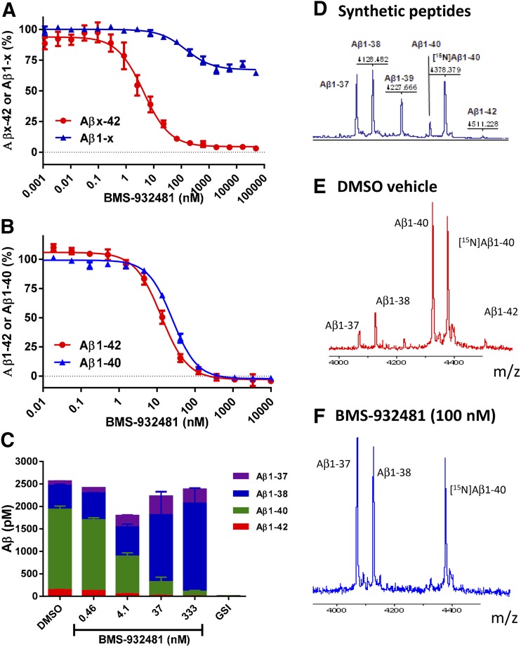
Fig. 1.
In vitro activity of BMS-932481. (A) H4-APPsw cell cultures were incubated overnight with BMS-932481 at a range of concentrations from 1 pM to 50 μM. Aβx-42 and Aβ1-x concentrations were determined simultaneously using the automated multiplex homogeneous time-resolved fluorescence assays. Error bars show standard error for seven independent assays. IC50 values (or inhibition percentage) with standard deviations are as follows: Aβx-42 IC50 = 5.5 ± 3.6 nM; Aβ1-x maximum inhibition = 30 ± 10% at 50 μM. (B) H4-APPsw cell cultures were incubated overnight with BMS-932481 at a range of concentrations from 10 pM to 10 μM. Aβ1-42 and Aβ1-40 were determined using automated homogeneous time-resolved fluorescence assays. Error bars show standard error for three independent assays in which both Aβ1-42 and Aβ1-40 were determined in parallel from the same cell cultures. IC50 values with standard deviations are as follows: Aβ1-42 IC50 = 6.6 ± 2.3 nM; Aβ1-40 IC50 = 25 ± 7.9 nM. (C) H4-APPsw cell cultures were incubated overnight with BMS-932481 at a range of concentrations from 0.46 to 333 nM, 0.1% dimethylsulfoxide (DMSO) vehicle, or GSI BMS-299897 at a concentration of 1 μM. Concentrations were determined for Aβ1-42, Aβ1-40, Aβ1-38, and Aβ1-37 using the 4-plex Meso Scale Diagnostics assays. Error bars indicate standard error for two replicate wells. (D) An equimolar mix of synthetic peptides, including [14N]Aβ1-40, was evaluated by matrix-assisted laser desorption/ionization mass spectroscopy. (E and F) H4-APPsw cell cultures were incubated overnight with 0.1% dimethylsulfoxide (DMSO) (E) or BMS-932481 (F) at a concentration of 100 nM, then Aβ peptides were immunoprecipitated and evaluated by matrix-assisted laser desorption/ionization mass spectroscopy.

