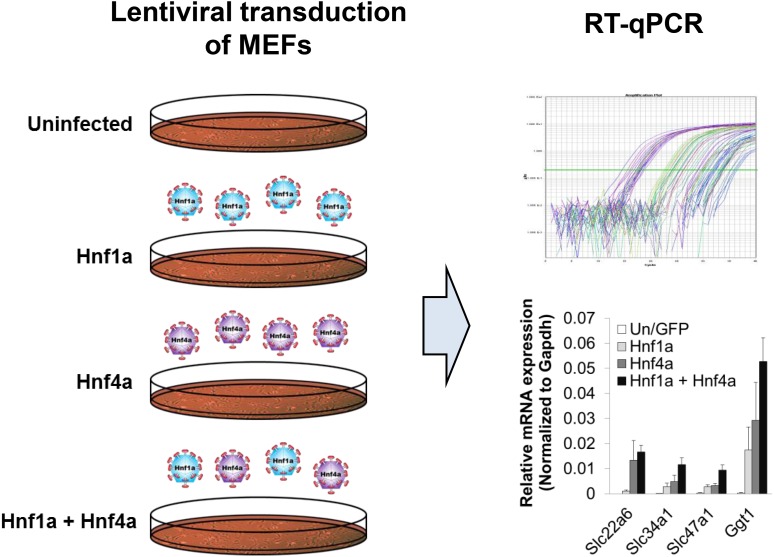
Fig. 1.
Lentiviral transduction of MEFs followed by quantitative reverse-transcription PCR (RT-qPCR) determination of changes in gene expression for selected PT markers. (Left panel) MEFs isolated from different days of gestation (i.e., E13.5, E15.5, and E16.5) were transduced without (uninfected) or with lentivirus expressing either GFP or the transcription factors Hnf1a and Hnf4a (either singly or in combination). (Right panel) The expression of several markers of the kidney proximal tubule [i.e., transmembrane transporters, Slc22a6 (Oat1), Slc34a1 (NaPi-2a), and Slc47a1 (Mate1), as well as the PT brush border marker Ggt1] was determined by quantitative reverse-transcription PCR. The graph on the bottom right shows the induction of several of these markers upon transduction of the MEFs with Hnf1a and Hnf4a; mean ± S.E.M. (n = 3) (Supplemental Fig. 1). Gapdh, glyceraldehyde-3-phosphate dehydrogenase.

