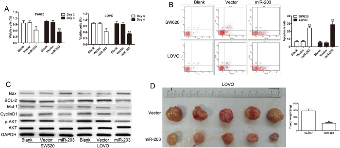
Figure 3. miR-203 inhibited cell growth and tumor growth.
(A) The proliferation rate of the miR-203 and the vector transfected cell lines as determined by the Cell Proliferation Assay Kit. (B) Cell apoptosis in SW620 and LOVO cells by flow cytometric assay. (C) The expression levels of Mcl-1, Bcl-2, cyclin D1, Bax, p-AKT and AKT were detected in the SW620 and LOVO cells transfected with the control or miR-203. (D) Representative images of the LOVO cell tumors expressing miR-203 or the vector. A quantitative analysis of the weight of the subcutaneous tumors from the two groups. *p < 0.05, **p < 0.01.

