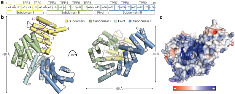
Figure 1. Structural overview of human IFIT5.
a, Secondary structure, TPR motif and subdomain organization of IFIT5. b, Orthogonal views of IFIT5 with helices represented as cylinders. c, Surface representation of IFIT5 coloured by electrostatic potential (using APBS) from negative ( −5kTe−1; red) to positive (+5kTe−1; blue).

