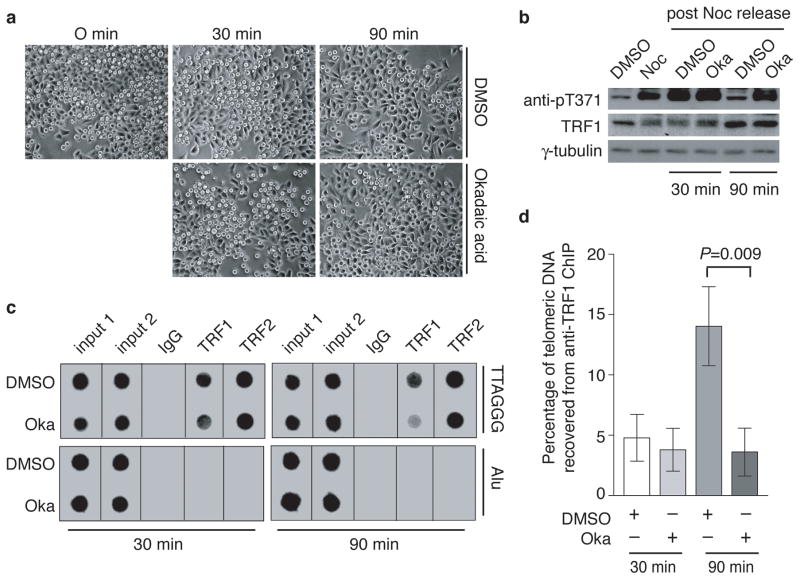
Figure 3. T371 of TRF1 dephosphorylated in late mitosis and this dephosphorylation allows the re-association of TRF1 with telomeric DNA.
(a) Live cell images for HeLaI.2.11 cells released from a nocodazole arrest into fresh media containing either DMSO or okadaic acid (1 μM) for 0–90 min. (b) Western analysis performed with anti-pT371, anti-TRF1 or anti-γ-tubulin antibody. (c) Dot blots of ChIPs performed with anti-TRF1 or anti-TRF2 antibody. Nocodazole-arrested HeLaI.2.11 cells were released into fresh media containing either DMSO or okadaic acid (Oka) for either 30 min or 90 min. (d) Quantification of anti-TRF1 ChIPs from (c). The P value was calculated using a Student’s two-tailed t test. Standard deviations derived from at least three independent experiments are indicated.

