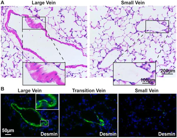
Figure 2. Only the large pulmonary veins contain cardiomyocytes.
(A) H&E-stained large and small pulmonary veins. Large vein inset shows region containing morphologically characteristic cardiac cells. Small vein inset shows absence of this layer. (B) Immunofluorescence image of large and small pulmonary veins stained with the intermediate filament protein desmin (green), marking cardiomyocyte striations (inset, double-arrow) and demonstrating the loss of cardiac cells in small veins. Nuclei are stained blue with Hoechst dye.

