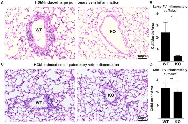
Figure 4. αT-cat loss decreases large, but not small pulmonary vein inflammation.
H&E-stained large (A) and small (C) PVs of both WT and αT-cat KO mice exposed to HDM. (B & D) Inflammatory cuff size was measured by dividing the area by the thickness of the muscle (large vein) or lumen size (small vein). AW=airway, PA=Pulmonary artery, PV=Pulmonary vein. *p<0.05, Student’s t-test, n=5. Error bars=S.E.M.

