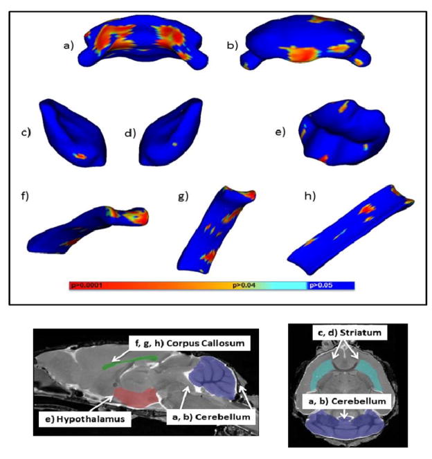
Fig. 5.
The effects of GD8 alcohol exposure on regional brain shapes. Top panel. Areas portrayed in blue are statistically unchanged from vehicle, while areas in other colors are significantly different between alcohol and vehicle treated mice. a and b) portray the anterior and posterior views of the cerebellum. c and d) portray the left and right striatum. e) portrays the anterior view of the hypothalamus. f, g, and h) portray the anterior, ventral, and dorsal views of the corpus callosum The colored bars indicate levels of significance. Bottom panel. Parasagittal and horizontal MRI images of a PD 45 mouse brain highlighting regions in which significant shape changes had occurred.

