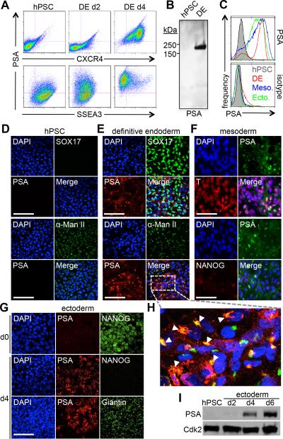
Figure 1.
Polysialic acid is expressed upon differentiation to all three germ layers. (A) Flow cytometry of hPSC differentiation to definitive endoderm showing surface expression of PSA along with endoderm marker CXCR4 (Top) and pluripotency marker SSEA3 (Bottom). (B) Immunoblot of PSA expression in hPSC and DE. (C) Flow cytometry of PSA expression (Top) in hPSC (Shaded gray), DE (Red), mesoderm (Blue), and NCC (Green). PSA+ fractions in each cell type are 2.9%, 79.7%, 72.4% and 81.1%, respectively. Isotype control is shown for comparison (Bottom). (D-H) Immunostaining of PSA expression in hPSC differentiation. Scale bar, 100μm. (D) hPSC: PSA and DE marker SOX17 (Top), PSA and Golgi marker α-mannosidase II (Bottom). (E) DE: PSA and SOX17 (Top), PSA and α-mannosidase II (Bottom). (F) Mesoderm: PSA and T (Top), PSA and NANOG (Bottom). (G) NCC: PSA and NANOG (Top), PSA and Golgi marker Giantin (Bottom). (H) Zoom of PSA and α-mannosidase II in DE. Arrows point to overlap of PSA and α-mannosidase II. (I) Time course immunoblot of PSA expression during hPSC differentiation to NCC (Top). CDK2 shown as loading control (Bottom). Abbreviations: hPSC, human pluripotent stem cell; DE, definitive endoderm; NCC, neural crest cell.

