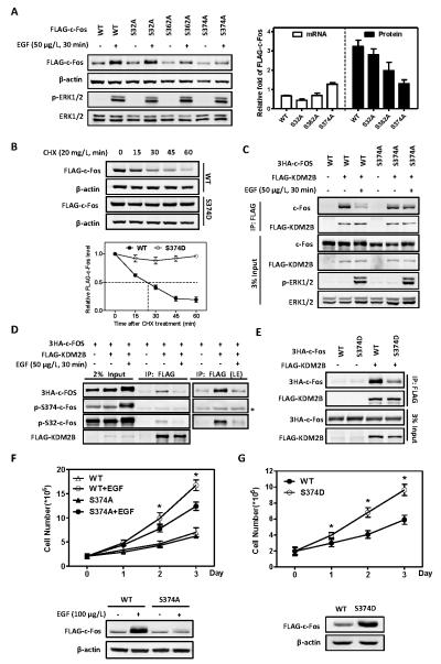
Figure 4. EGF-mediated phosphorylation at S374 stabilizes c-Fos and promotes cell proliferation.
(A) Mutation of S374 site to alanine abolishes EGF-induced c-Fos protein accumulation. HEK293 cells stably expressing FLAG-tagged c-Fos were deprived of serum for 8 h, and then treated with EGF (50 μg/L) for 30 min. The protein and mRNA levels of FLAG-c-Fos were determined by western blot and qRT-PCR, respectively, and normalized against β-actin. Data are shown as relative fold change over cells without EGF treatment. Error bars represent ±SD for duplicate experiments.
(B) Mutation of S374 site to aspartic acid renders c-Fos resistant to degradation. HEK293 cells stably expressing FLAG-tagged c-Fos WT or S374D mutant were treated with 20mg/L CHX for the indicated length of time. The half-life of c-Fos protein levels were determined by western blot and normalized against β-actin (Bottom). Error bars represent ±SD for triplicate experiments.
(C) The reduction of c-Fos and KDM2B interaction by EGF is inhibited by c-Fos S374A mutant. HEK293 cells were co-transfected with plasmids expressing indicated proteins and then treated with or without EGF (50 μg/L) for 30 min. The interactions between c-Fos and KDM2B were determined by Co-IP.
(D) KDM2B preferentially interacts with S374 non-phosphorylatable c-Fos. HEK293 cells were co-transfected with plasmids expressing the indicated proteins and then treated with or without EGF (50 μg/L) for 30min. The FLAG-KDM2B was immunoprecipitated and western blot was performed to detect the co-precipitated c-Fos with indicated antibodies. * indicates the heavy chain background around 55 KDa.
(E) S374D mutation of c-Fos hinders its binding to KDM2B. HEK293 cells were co-transfected with plasmids expressing indicated proteins, and the interactions between c-Fos and KDM2B were determined by Co-IP.
(F) EGF-induced cell proliferation is compromised by S374A mutant of c-Fos. HEK293 cells stably expressing FLAG-c-Fos WT or S374A mutant were cultured in the absence of serum and treated with or without EGF (100 μg/L) for 0, 1, 2 or 3 days, as indicated. EGF was replenished every day. Cell numbers were counted each day. Western blot was performed to show FLAG-c-Fos protein levels on the 3rd day. * denotes the p < 0.05 for cells stably expressing S374A mutant versus wild-type c-Fos under EGF treatment. Error bars represent ±SD for triplicate experiments.
(G) c-Fos S374D mutant is more potent than wild-type in promoting cell proliferation. HEK293 cells stably expressing FLAG-c-Fos wild-type or S374D mutant were cultured in the absence of serum for 0, 1, 2 or 3 days, as indicated. Cell numbers were counted each day. Western blot was performed to show FLAG-c-Fos protein levels on the 3rd day. * denotes the p < 0.05 for cells stably expressing S374D mutant versus wild-type c-Fos. Error bars represent ±SD for triplicate experiments.

