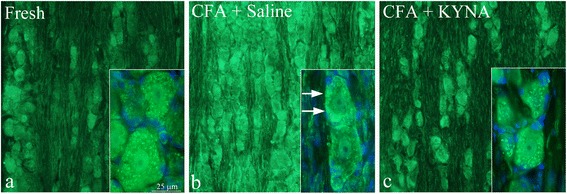
Fig. 2.

IL-1β in the trigeminal ganglion. a IL-1β immunoreactivity was observed in the neuronal cytoplasm (in a granular manner), in a few nuclei and in the nerve fibers of fresh animals. No immunoreactivity was detected in the SGC. b After i.p. treatment with saline for 7 days, increased IL-1β immunoreactivity was observed both intracellularly in the neurons, and in the fibers. In addition, a “ring” IL-1β immunoreactivity close to the neuronal cell membrane was evident. c Following i.p. treatment with KYNA for 7 days, the homogenous immunoreactivity close to the cell membrane disappeared, returning to the granular cytoplasmatic pattern observed in fresh animals. No difference could be noted in the neuronal nuclei and in fibers, and no immunoreactivity was detected in the SGC
