Figure 1.
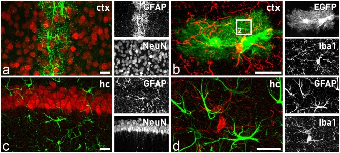
Astrocytes and microglia in the forebrain. Distinct subtypes of astrocytes are present throughout the brain, while microglial cells seem to be more homogenously distributed. In the cortex only astrocytes in close contact to blood capillaries express significant levels of GFAP (A), while all astrocytes are closely intermingled with adjacent neurons (A) or microglia (B). In contrast, in the hippocampus all astrocytes express GFAP (C). They are also in close contact to neurons (B) and microglial cells (D). Comparison of GFAP staining (D) with EGFP expression in (B) of TgN (GFAP-EGFP)GFEC transgenic mice reveals only in the latter the fine arborization of perisynaptic and perivascular astrocytic processes. The square in (B) indicates the magnified view that is schematically depicted in Figure 2. Scale bars indicate 20 μm.

