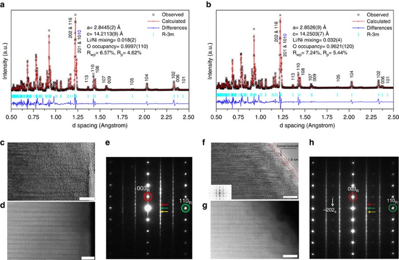
Figure 2. Structural characterizations of the pristine and GSIR LR-NCM.
(a,b) TOF ND patterns for the pristine and GSIR LR-NCM. On the basis of these results, the oxygen vacancy for the GSIR LR-NCM is around three per cent higher than that of the pristine LR-NCM sample; (c) high-resolution transmission electron microscopy (HRTEM) image and, (d) high-angle annular dark-field—STEM (HAADF-STEM) image for the pristine LR-NCM; (e) electron-diffraction (ED) pattern for the pristine LR-NCM. The elongation of these spots are along [001]R* as well as [001]M* direction. The spots indicated with red, green and yellow arrows represent (110), (020) and (−110) of the monoclinic Li2MnO3 (donated as M) variants [1–10]M, [100]M and [110]M, respectively. The spot marked by a red circle is shared by (003)R of the rhombohedral (donated as R), as well as the structure (001)M of all monoclinic variants. The spot marked by a green circle is shared by (110)R of the rhombohedral, as well as the structure (33-1)M, (060)M and (–331)M of the monoclinic. (f) HRTEM image with fast Fourier transform (FFT) about the surface and (g). HAADF-STEM image for the GSIR LR-NCM; (h) ED for the GSIR LR-NCM. The weak spot indicated by the white arrow can only be indexed as the structure (−202)S of the spinel (donated as S). Scale bars, 5 nm.

