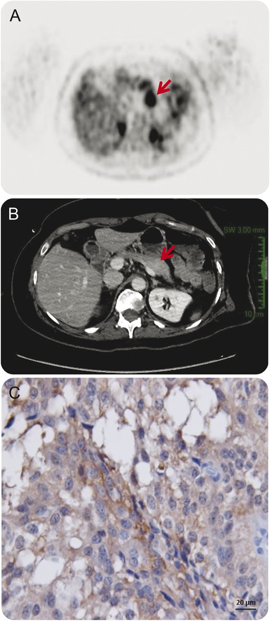
Figure. FDG-PET and CT scans of the patient with pancreatic tumor.

(A) The FDG-PET scan revealed a 4-cm-diameter tumor localized in the pancreatic body (arrow), (B) underdiagnosed on previous abdominal CT scan (arrow). (C) The pancreatic tumor of the patient immunolabeled with a specific antibody for the GluN1 subunit of the NMDA receptor (original magnification, ×400). FDG = [18F]-fluorodeoxyglucose.
