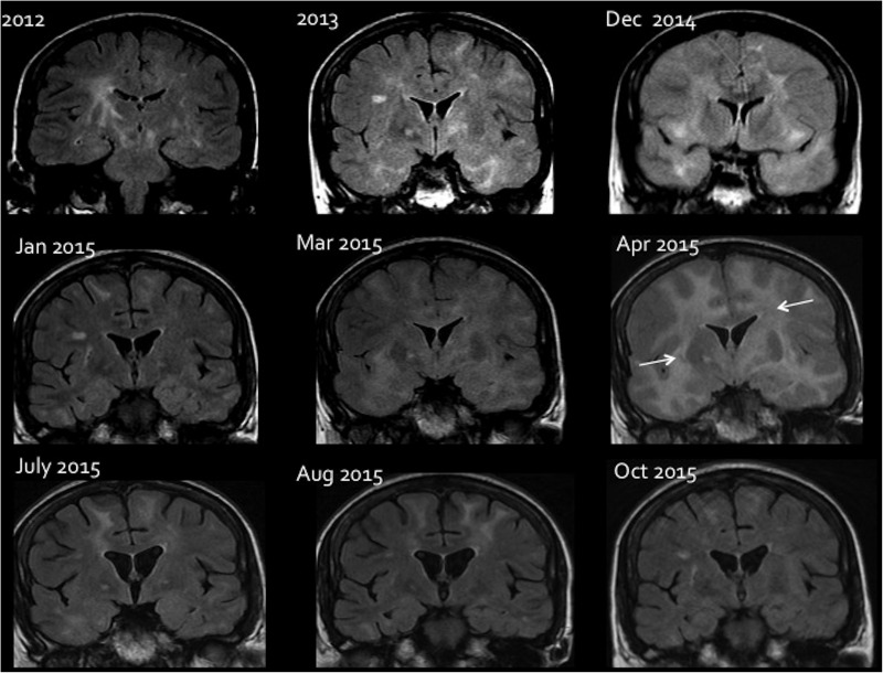
Figure 1.
Sequential MRI changes during treatment of CD8+E. Coronal FLAIR images showing the brain parenchymal changes and response to different treatment. In 2012, an abnormal high signal is seen in the basal ganglia bilaterally with signal change in the white matter in keeping with encephalitis. Image taken from 2013 shows a marked worsening of white matter signal change more extensive within the temporal lobes. In 2014, there is a bilateral white matter signal change with diffuse brain swelling and effacement of all cortical sulci. Following steroid initiation in December 2014, there is significant improvement in the signal change as well as mass effect on the January 2015 image. A worsening in March 2015 and a more pronounced deterioration in April 2015 (arrows), after reduction in steroids. A subsequent increase in steroids shows a remarkable improvement in July 2015. The patient was later started on mycophenolate mofetil in July 2015; this allowed a further amelioration of lesions as seen in August 2015 and October 2015, despite a tapering of steroids.

