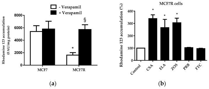
Figure 2.
Accumulation of rhodamine 123 in MCF7 and MCF7R cells. (a) Cells were exposed to 5.25 µM rhodamine 123 for 30 min at 37 °C in the absence or presence of 50 µM verapamil. After washing, rhodamine 123 accumulation was quantified by spectrofluorimetry. Data are expressed as fluorescence arbitrary unit (FAU)/mg protein and are the means ± SEM of three independent experiments. *, p < 0.05 when compared to MCF7 cells; §, p < 0.05 when compared to counterparts not exposed to verapamil. (b) MCF7R cells were exposed to 5.25 µM rhodamine 123 for 30 min at 37 °C in the absence (control) or presence of 100 µM cyclosporin A (CSA), 1 µM elacridar (ELA), 1 µM zosuquidar (ZOS), 2 mM probenecid (PRB) or 10 µM fumitremorgin C (FTC). After washing, cellular rhodamine 123 accumulation was quantified by spectrofluorimetry. Data are expressed as % of fluorescent dye accumulation in control MCF7R cells exposed only to rhodamine 123, arbitrarily set at 100%, and are the means ± SEM of three independent assays. *, p < 0.05 when compared to control cells.

