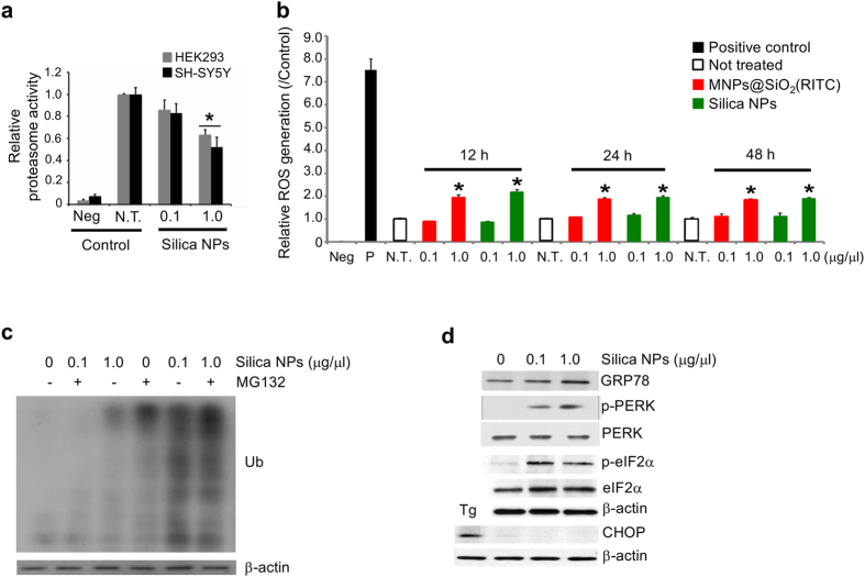
Figure 3. Generation of intracellular effects after treatment with MNPs@SiO2(RITC) and silica NPs.
(a) Inhibition of proteasome activity in HEK293 and SH-SY5Y cells treated with silica NPs. A luminescence-based assay was performed on untreated, 0.1 μg/μl, and 1.0 μg/μl silica NPs-treated HEK293. (b) ROS generation in MNPs@SiO2(RITC)- and silica NPs-treated HEK293 cells. Cells were treated with 0.1 μg/μl or 1.0 μg/μl of MNPs@SiO2(RITC) or silica NPs for 12, 24, and 48 h, followed by fluorescence measurement after staining with DCFH-DA. Intensities were normalized relative to non-treated control. One mM of hydrogen peroxide (H2O2) was used as positive control and media only was used as negative control. Data represent means values ± S.D. of triplicate measurements relative to the non-treated control. (c) Accumulation of ubiquitinated proteins in silica NPs-treated cells. HEK293 cells were treated with 0.1 μg/μl and 1.0 μg/μl of silica NPs for 36 h, followed by treatment with MG132 or vehicle for 12 h. (d) ER stress-mediated apoptosis did not occur in silica NPs-treated cells. HEK293 cells were treated with 0.1 μg/μl or 1.0 μg/μl of silica NPs for 12 h, and the ER stress-related proteins GRP78, phosphorylated PERK (p-PERK), PERK, phosphorylated eIF2α (p-eIF2α), eIF2α, and CHOP were analyzed by western blotting. Ten μM Thapsigargin (Tg) was used as positive control. β-actin was used as loading control.

