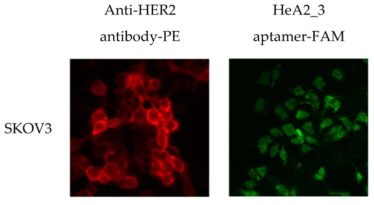
Figure 7.
Fluorescence microscopy analysis of internalization of aptamer HeA2_3 into SKOV3 cells. Cultured SKOV3 cells were stained with anti-HER2 antibody (red) or HeA2_3 aptamer (green) and internalization was evaluated by fluorescence microscopy. Images were taken at a magnification of 400×. All experiments were performed at 37 °C as lower temperatures may inhibit internalization.

