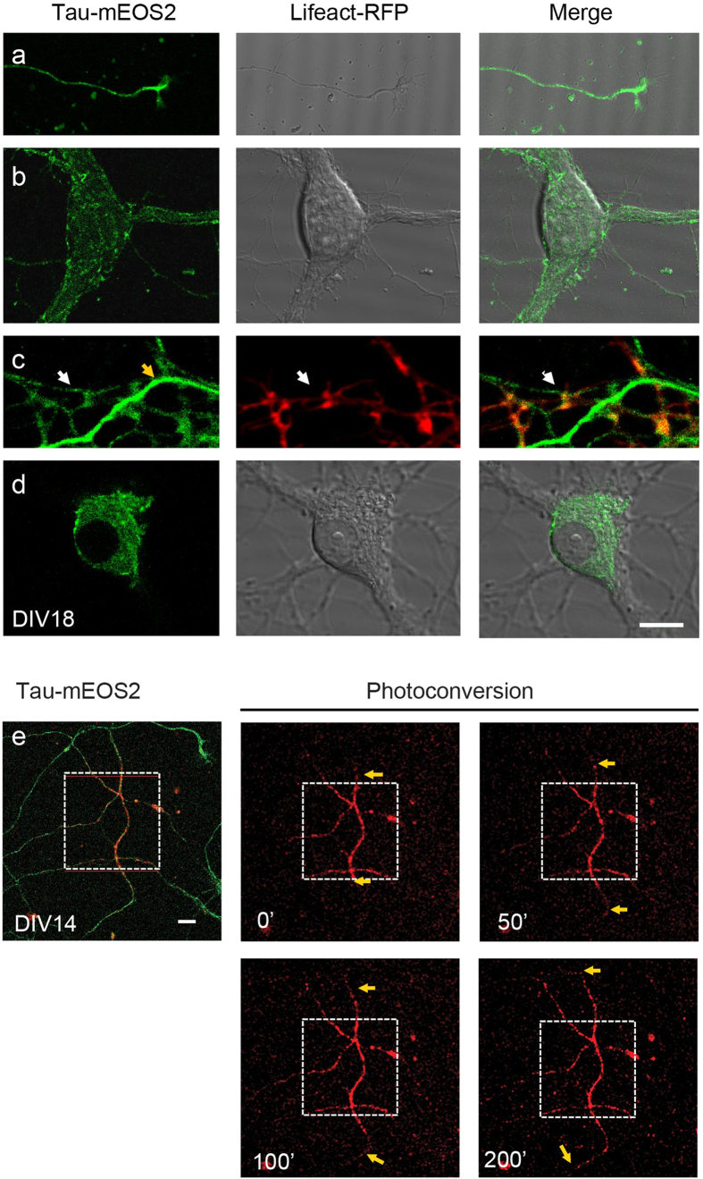
Figure 2. Tau-mEOS2 is localized to axons, dendrites and the soma but excluded from the nucleus.
Mature DIV18 hippocampal cultures from Tau-mEOS2 mice cotransfected with the spine marker Lifeact-RFP to reveal dendritic spines. (a) Tau-mEOS2 mice show a strong axonal expression of EOS-tagged Tau, with a gradient towards the growth cone of distal axons. (b) Tau-mEOS2 is also present in the somatodendritic domain, (c) with very low levels in dendritic spines (zoom-in, white arrow: dendrite; yellow arrow: axon). (d) Within the limit of detection, EOS-tagged endogenous Tau cannot be visualized in the nucleus. (e) Time-lapse imaging of photo-converted mEOS2-tagged Tau reveals bidirectional transport of Tau in the axon at about ~0.25 μm/s. Yellow arrows indicate how far photo-converted Tau travelled. Scale bar: 10 μm (a–d) and 15 μm (e).

