Figure 1.
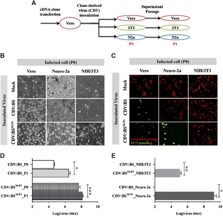
Enabling EV71 infection of mouse cell lines by replacing the EV71:BS capsid with EV71:TLLm capsid sequences. (A) Schematic representation of the cell infection experiments. 3T3, NIH/3T3; N2a, Neuro-2a; P0, passage 0; P1, passage 1. (B–E) Assessment of cellular infection induced by CDVs at 48 h p.i. Cells inoculated with CDVs at an MOI of 1 were observed for evidence of lytic CPE (B) and expression of viral proteins (C). Production of viable viral progeny was also determined by measuring viral titers from inoculated Vero (D), NIH/3T3 and Neuro-2a cells (E). Live cell images were taken at × 40 magnification and are shown in grayscale. Fluorescence images were taken at × 40 magnification and are shown with enhanced contrast. Viral antigens were labeled with FITC (green), and cells were counterstained with Evans blue (red). Titers from Vero cells were measured at P0 and P1, while titers from NIH/3T3 and Neuro-2a cells were measured at P0. Titrations were performed with dilutions of 10−1 to 10−10 in Vero cells following viral disaggregation with chloroform. The results are representative of two independent studies. For D and E, a modified t-test with Welch's correction for unequal variance was used to compare mean values (n=4). Error bars represent the s.d. *P<0.05; **P<0.005, ***P<0.0005. clone-derived virus, CDV; cytopathic effect, CPE; multiplicity of infection, MOI.

