Figure 5.
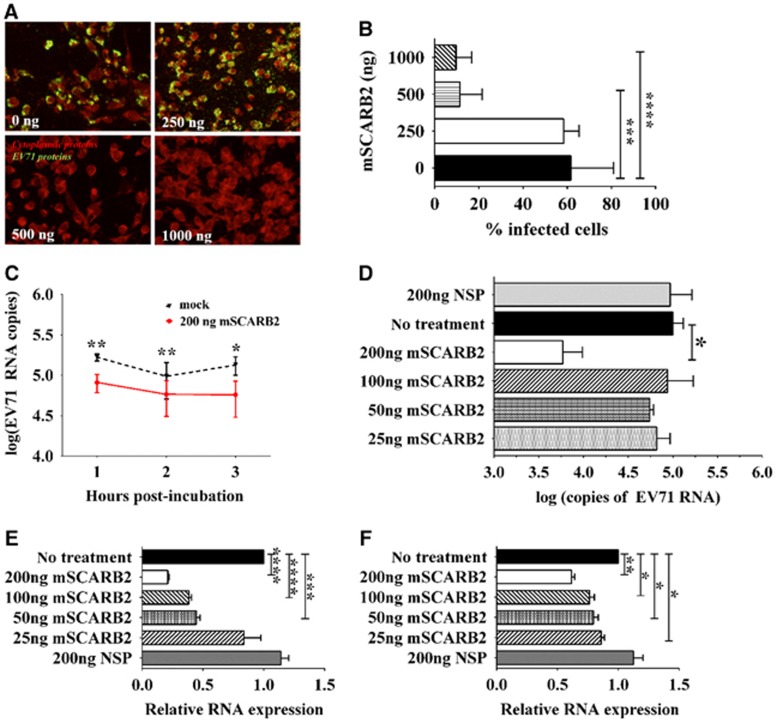
Viral competitive entry/uncoating assays by pre-incubation of EV71 with the murine SCARB2 (mSCARB2) protein. (A, B) Cellular infection of NIH/3T3 cells with EV71:TLLmv pre-incubated with various concentrations of soluble mSCARB2 protein prior to viral inoculation. Viral antigens were detected by fluorescence immunostaining at 48 h p.i. (A). Fluorescence images were taken at × 40 magnification and are shown with enhanced contrast. Viral antigens were labeled with FITC (green), and cells were counterstained with Evans blue (red). The number of infected cells was counted in ten independent fields of view (B). (C, D) Viral uncoating study, where 106 CCID50 of EV71:TLLmv was incubated with various amounts of soluble mSCARB2 protein. Viral RNA copies were absolutely quantified after incubation with 200 ng of mSCARB2 for 3 h (C) or after incubation with various concentrations of mSCARB2 for 2 h (D). (E, F) Effect of virus pre-incubation with soluble mSCARB2 on cellular infectivity. Relative quantitation of EV71 RNA in total cellular RNA extracted from NIH/3T3 cells inoculated with either EV71:TLLmv (E) or EV71:TLLm (F) pre-incubated with soluble mSCARB2 at an MOI of 10. Total cellular RNA was extracted at 2 h p.i., and viral RNA was quantified by the ΔΔCT method using β-actin as an internal control. For B, a Mann–Whitney U-test was used to compare medians (n=8). Error bars represent the range. For C–F, a t-test with Welch's correction for unequal variance was used to compare mean values (n=4). Error bars represent the s.d. *P<0.05, **P<0.005, ***P<0.0005, ****P<0.0001. cell culture infective dose, CCID; non-specific protein, NSP.

