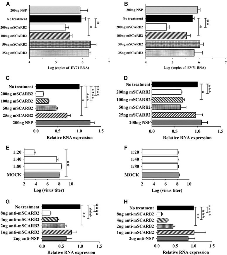
Figure 7.
Assessing the role of the mSCARB2 protein in CDV:BSM-P1 and CDV:BS-VP1K98E,E145A,L169F infection of murine cells. (A–D) Pre-incubation of 106 CCID50 CDVs with the mSCARB2 protein for in vitro uncoating (A, B) or for cellular infection studies of NIH/3T3 cells (C, D). The viral RNA in the samples was extracted and quantified by RT–PCR (qRT–PCR). (E–H) Blocking viral entry by incubating NIH/3T3 cells with anti-mSCARB2 sera prior to inoculation with virus at an MOI of 10. Infection was assessed by determining viral titers in culture supernatant with dilutions of 10-1 to 10−10 in Vero cells at three days p.i. following chloroform viral disaggregation (E, F), and relative quantitation of EV71 RNA in extracted total cellular RNA by the ΔΔCT method using β-actin as an internal control (G, H). Tests were separately performed for CDV:BSM-P1 (A, C, E, H) and CDV:BS-VP1K98E,E145A,L169F (B, D, F, G). For A–H, a t-test with Welch's correction for unequal variance was used to compare mean values (n=4). Error bars represent the s.d.; *P<0.05, **P<0.005, ***P<0.0005, ****P<0.0001. clone-derived virus, CDV; multiplicity of infection, MOI; quantitative reverse transcription–PCR, qRT–PCR.

