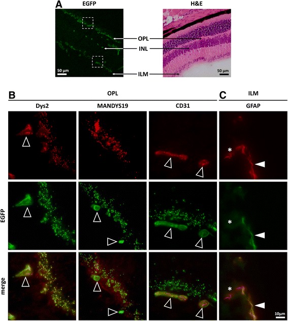
Fig. 6.

Dystrophin-EGFP expression in the retina. a Presence of the natural EGFP signal in the different layers of the retina: outer plexiform layer (OPL), inner nuclear layer (INL), and inner limiting membrane (ILM) (left image). These regions are depicted on a corresponding H&E stained cross section from the retina of a wild-type mouse (right image). A higher magnification of the EGFP-positive regions highlighted by the squares in a is presented in b and c. b Immunofluorescent staining with antibodies against dystrophin (Dys2, MANDYS19, red) and CD31 (red) showed differential colocalization of natural EGFP signal (green) in the retinal OPL of Dmd EGFP mice. Arrows indicate EGFP-positive retinal blood vessels that were detected with Dys2 and anti-CD31 antibody, but not with the rod-specific antibody. Strong punctuated expression of the natural EGFP was observed in the OPL region corresponding to the photoreceptor terminals. In this region, an exact superposition with the Dys2 signal and some overlay with the MANDYS19 signal were observed, whereas no anti-CD31 signal was detected. c Immunofluorescent staining with antibodies against the glial fibrillary protein (GFAP, red) showed partial colocalization with the natural EGFP signal at the ILM. Arrowheads indicate colocalization between GFAP and EGFP at the glial endfeet. The asterisk marks the GFAP positive region, which did not express EGFP, corresponding to the glial cell body
