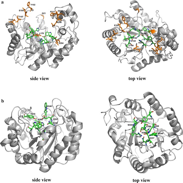
Fig. 8.

a The predicted 3D structure of cel7482 (side view and top view) using the crystal structure of a family 5 endoglucanase (PDB: 1ceo) as modeling template. The colored sticks represent the conserved residues in the active site. The orange sticks represent the different residues between cel7482 and cel3623. b The predicted 3D structure of cel36 (side view and top view) using the crystal structure of endo-1,4-β-glucanase from Bacillus subtilis 168 (PDB: 3pzt) as modeling template. The colored sticks represent the conserved residues in the active site
