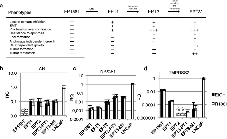
Fig. 2.

AR is expressed in a mesenchymal context, but target genes are repressed. a An experimental model of stepwise transformation of prostate cells to malignant cells. The model was started from benign EP156T epithelial cells obtained during surgery. The cells were grown to confluence and kept for almost 4 months without splitting to select for cells with reduced cell-to-cell contact inhibition. EPT1 cells appeared following EMT of EP156T cells. EPT1 cells were grown to confluence for several weeks and foci appeared in the monolayers. EPT2 cells were picked from the foci and selected and cloned by growth in soft agar. Neither EP156T nor EPT1 were able to grow in soft agar. Individual clones of EPT2 were next grown in protein free medium, and the selected cells were tumorigenic and generated EPT3 cells which were recovered from subcutaneous mice tumors and transduced with a GFP-luciferase vector [15].*Orthotopic injection of EPT3-GFP-luc cells in mice resulted in the EPT3-PT1 cells derived from the primary tumor. EPT3-M1 cells were isolated from abdominal metastasis. The accumulation of malignant features as one cell type was derived from its progenitor is listed. RT-qPCR of b AR, c NKX3-1 and d TMPRSS2 expression in epithelial (EP156T) and derived mesenchymal cells compared to LNCaP, treated with 10 nM R1881. Error bars show 95 % confidence intervals. N.D. = not detected. RQ = relative quantity
