
Fig. 3.
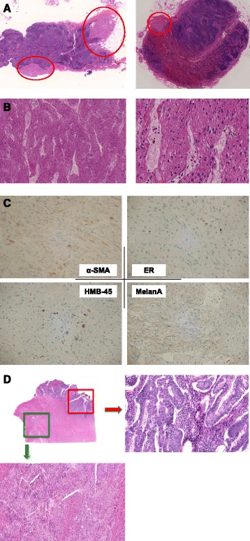
a The maximum size lymph node was dissected from the left lateral aortic lesion. Microscopically, masses was composed of neoplastic smooth muscle leiomyoma-like tumor cells with clear to eosinophillic cytoplasm arranged in alveolar pattern without necrosis. b Slit-like vascular channels lined by endothelial cells were seen in the lymph node. The cells with irregular shaped nucleus were arranged in nested pattern. c Lymph node tumors were focally positive for smooth muscle actin (SMA), caldesmon, Melan A, HMB-45, and ER, characteristics suggestive of LAM. Estrogen receptor (ER) was also focally positive. d Spindle cell proliferation suggestive of regional LAM tissue was seen in the re-examination of uterine tissue from the primary operation