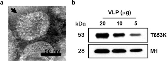
Fig. 2.

Characterization of virus-like particles (VLPs). Electron microscopy and VLP size determination. Negative staining of VLPs was performed followed by transmission electron microscopy (TEM). The size is between 40 and 120 nm (a). Western blot analysis. VLPs (20, 10, 5 μg) were loaded for SDS-PAGE. Polyclonal mouse anti-T. spiralis antibody was used to probe T653k protein and anti-M1 monoclonal antibody was used to determine M1 protein (b)
