
Figure 2.
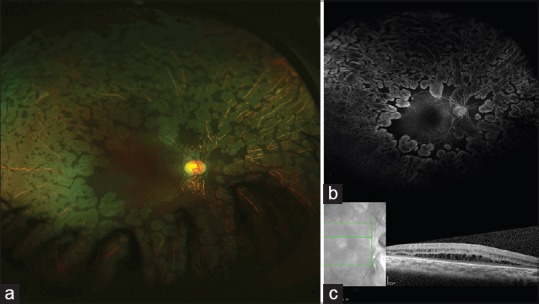
(a) Ultrawide field fundus image of 7-year-old girl (Case 2) shows multiple distinct focal areas of chorioretinal atrophy suggestive of gyrate atrophy of choroid and retina. (b) Ultrawide field fundus fluorescein angiogram did not show any leak at macula even in late phase. (c) Optical coherence tomogram showed hyporeflective spaces separated by multiple linear bridging elements suggestive of foveoschisis