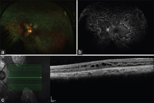
Figure 3.

(a) Ultrawide field fundus image of the left eye of 8-year-old boy (Case 3) shows multiple sharply demarcated lesions sparing macula, typical of gyrate atrophy of choroid and retina. (b) Ultrawide field fundus fluorescein angiogram did not show any leak at macula even in late phase. (c) Optical coherence tomogram showed multiple hyporeflective areas with vertical intervening bridges of retinal tissue suggestive of foveoschisis
