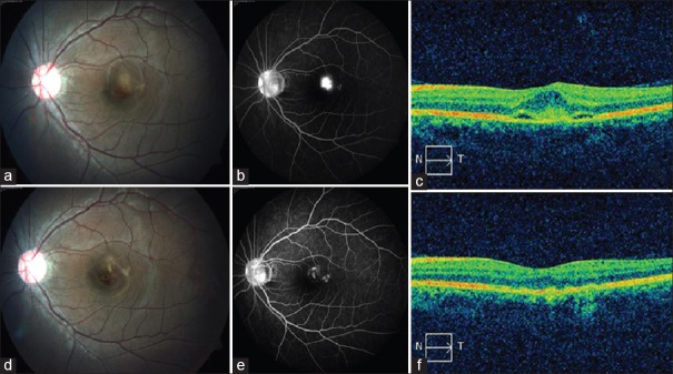
Figure 1.
(a) Fundus photograph shows juxtafoveal choroidal neovascular membrane with associated hemorrhage and subretinal fluid. (b) Fundus fluorescein angiography late phase shows intense leak from choroidal neovascularization with fuzzy borders. (c) Optical coherence tomography shows spindle-shaped hyper-reflective membrane between retinal pigment epithelium and neurosensory retina with subretinal fluid. (d) Fundus image 3 months postphotodynamic therapy and bevacizumab shows regression of choroidal neovascularization, resolution of retinal hemorrhage, and subretinal fluid. (e) Late phase fundus fluorescein angiography shows minimal staining of the scar with no leakage. (f) Optical coherence tomography demonstrates complete resolution of the subretinal fluid with normal foveal dip and regression of the membrane

