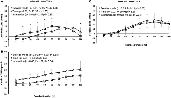
Figure 3.
(A–C) Cerebral oxygenation (COX) measured at the prefrontal cortex (PFC) during maximal controlled-pace (MIT) and self-paced (TT4km) exercise. Symbols indicate exercise mode (†) and time (‡) main effects, as well as time-by-exercise mode interaction effects (*). Panels (A–C) depict Δ[O2Hb], Δ[HHb], and Δ[THb].

