
Extended Data Figure 8. Infection of WT→RarHet or WT→WT chimeras with Murid herpesvirus-4.
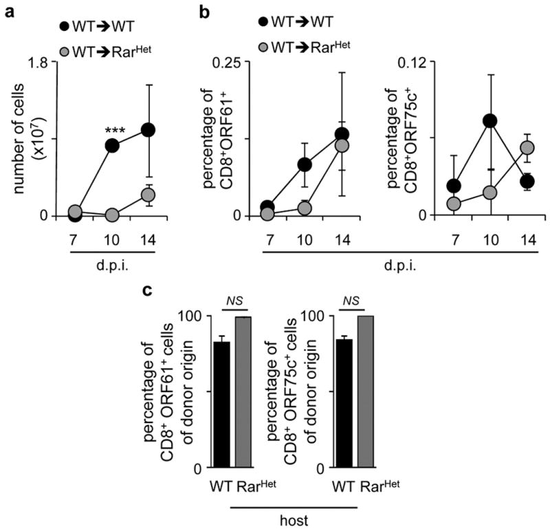
a, Intrathoracic lymph node cellularity in WT→WT and WT→RarHet chimeras at different days post infection (dpi). WT n=5; RarHet n=3. b, Percentage of CD8+ORF61+ (left) and ORF75c+ (right) T cells in intrathoracic lymph nodes at different days post infection (dpi). WT n=5; RarHet n=3. c. Percentage of donor CD45.1 CD8+ORF61+ (left) and CD8+ORF75c+ (right) T cells in WT→WT and WT→RarHet chimeras after infection with Murid herpesvirus-4. WT n=6; RarHet n=12. Error bars show s.e.. Two tailed t-test p values are indicated. *P<0.05; **P<0.01; ***P<0.001. n.s., not significant.