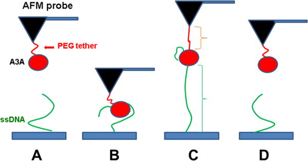
Figure 1.

Schematic representation of the experimental setup. Protein is covalently attached to the AFM probe via a PEG tether (red), and ssDNA (green) is covalently attached at the 5′ end to the functionalized mica surface. (A) Initial position, where A3A and ssDNA are far from forming a complex. (B) As the probe approaches the surface, A3A captures ssDNA and forms a complex. (C) Retraction step causing stretching of the PEG tether and ssDNA. (D) Rupture of the complex.
