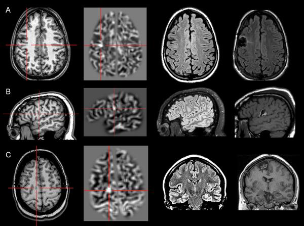
Figure 1.
Examples of three patients with MAP+ region; complete resection of the MAP+ region rendered all three patients seizure-free (>12 months). In Figures 1, 2 and 4, the crosshairs pinpoint the location of MAP+ region. First column: T1-weighted MPRAGE images used during pre-surgical evaluation. Second column: gray-white matter junction z-score file, as the output of MAP processing of the T1-weighted image shown in column one. Third column: T2-weighted FLAIR images, chosen to best depict the MAP+ region. Fourth column: post-surgical MRI indicating site and extent of resection. Pathology: A: FCD type IIA; B, FCD type IIB; C: FCD type IIA.

