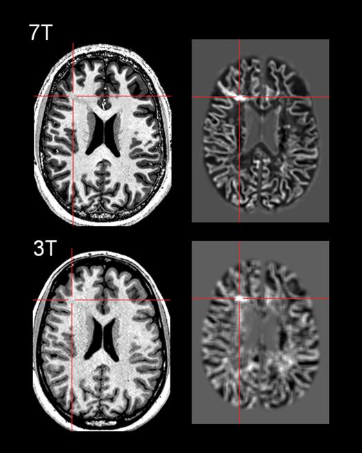
Figure 3.
Comparison of 7T and 3T images and MAP post-processing in the same patient with surgically confirmed FCD. Top row: 7T T1w MP2RAGE sequence, and the MAP gray-white junction feature map highlighting the subtle FCD with voxel size=0.5mm3. Bottom row: 3T T1w MPRAGE sequence, and the MAP gray-white junction feature map showing the same lesion, with voxel size=1mm3. The patient is a 21 year old right-handed male with intractable focal epilepsy of right frontal onset. Seizure semiology and ictal EEG (maximum evolvement in the right fronto-central region) were both concordant with location of the MRI lesion in the right middle frontal gyrus. The 3T MRI FCD lesion was further confirmed and re-illustrated by 7T MRI. MRI post-processing using MAP markedly enhanced visualization of blurring in the gray-white boundary. The patient underwent resection of the lesion guided by electrocorticography. FCD Type IA was found in surgical pathology.

