Fig. 4.
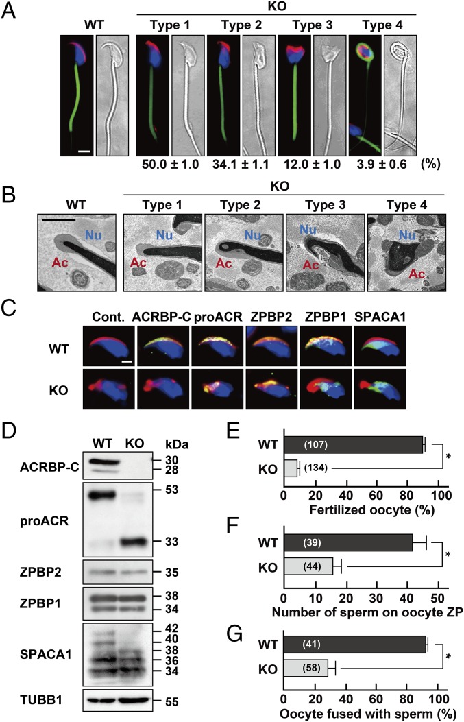
Characterization of ACRBP-deficient epididymal sperm. (A) Sperm morphology. The acrosome, mitochondria, and nucleus of cauda epididymal sperm from wild-type (WT) and ACRBP-deficient (KO) mice were stained with fluorescent-dye–labeled PNA (red), MitoTracker Green FM (green), and Hochest 33342 (blue), respectively. The KO sperm were morphologically divided into four types, and the rate of each cell type was determined. (Scale bar, 4.0 μm.) (B) TEM analysis. Ac, acrosome; Nu, nucleus. (Scale bar, 1.0 μm.) (C) Immunostaining analysis. Sperm were immunostained with antibodies against proteins indicated (green) and also stained with fluorescent-dye–labeled PNA (red) and Hoechst 33342 (blue). No immunoreactive signal was detected when preimmune serum was used as the probe (Cont.). (Scale bar, 2.0 μm.) (D) Immunoblot analysis. Proteins were separated by SDS/PAGE and probed with antibodies against the sperm proteins indicated. (E–G) Functional assays of sperm. Capacitated sperm were subjected to assays for IVF (E), sperm/ZP binding (F), and sperm/oocyte fusion (G). The numbers in parentheses indicate those of the oocytes examined. All statistical significances are calculated using the Student t test. *P < 0.01.

