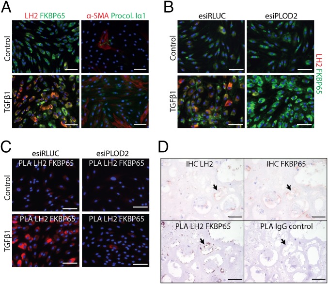
Fig. 1.
Endoplasmic reticulum-localized LH2 and FKBP65 exhibit physical interactions. (A) Immunofluorescent detection of LH2, FKBP65, aSMA, and procollagen-Iα1 in NHDFs with or without TGFβ1 treatment for 2 d. (B) Immunofluorescent detection of LH2 and FKBP65 in NHDFs transfected with esiRNA against PLOD2 or control (RLUC), followed by treatment with or without TGFβ1 for 2 d. (C) Fluorescent proximity ligation assay for LH2 and FKBP65 in NHDFs transfected with esiRNA against PLOD2 or control (RLUC), followed by treatment with or without TGFβ1 for 2 d. (D, Upper) Immunohistochemistry for LH2 and FKBP65 on serial sections from fibrotic explanted human renal allograft tissue. (D, Lower) PLA on identical serial sections for LH2 and FKBP65 or IgG isotypes as a control. The arrow indicates colocalization of LH2 and FKBP65 in the serial sections. (Scale bars: white, 100 µm; black, 50 µm.)

