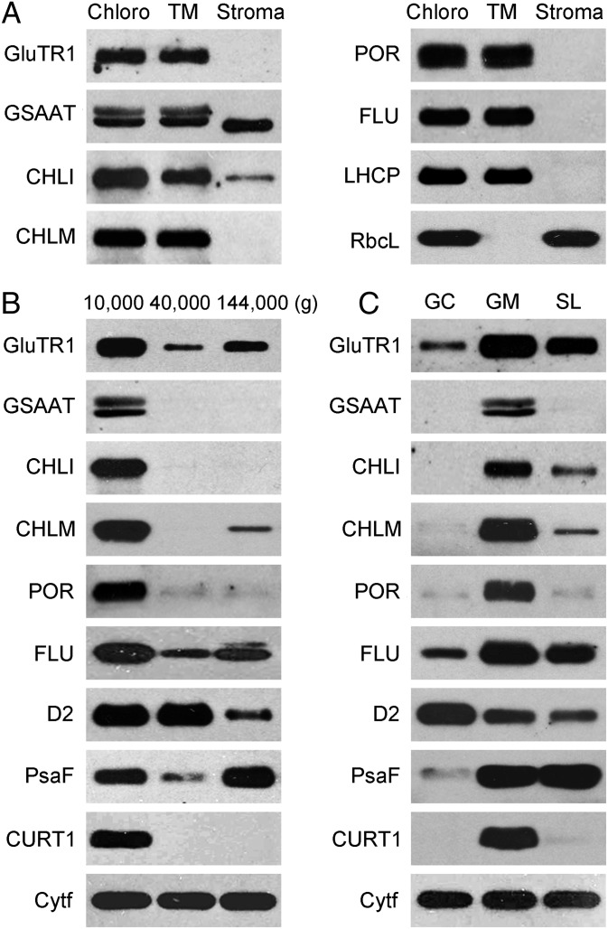
Fig. 5.
Enzymes of Chl biosynthesis and EX1 colocalize in grana margins. For the localization of enzymes catalyzing the initial and final steps of Chl synthesis, the same chloroplast fractions were analyzed as described in Fig. 1 C–E: (A) Chloroplasts (Chloro), thylakoid membranes (TM), and stroma (Stroma). (B) The three subfractions of chloroplast membranes that were pelleted at 10,000 × g, 40,000 × g, and 144,000 × g. (C) Grana core (GC), grana margins (GM), and stroma lamellae (SL). The localization of the following enzymes was determined by SDS/PAGE and immunoblotting: glutamyl-tRNA reductase (GluTR), glutamate-1-semialdehyde aminotransferase (GSAAT), Mg2+-chelatase subunit I (CHLI), Mg2+-protoporphyrin IX methyl transferase (CHLM), and PORs B and C. FLU, D2, PsaF, and CURT1 were used as marker proteins for the identification of the various chloroplast fractions. Cyt f was used as a loading control.

