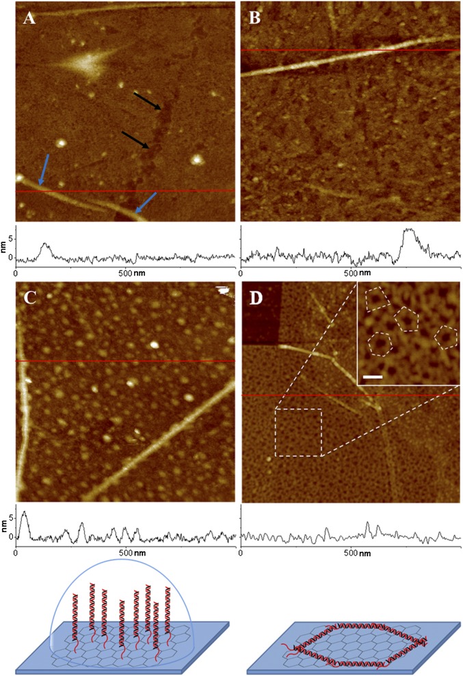
Fig. 3.
AFM images of graphene transistor surface with and without DNA strands. (A) Graphene surface in fluid is mostly flat with some defects (black arrows) and graphene wrinkles (blue arrows). (B) PASE-coated graphene surface in fluid showing a flat surface with a similar wrinkle height of 7 nm, as seen in A. (C) After binding of dsDNA on the PASE-coated graphene surface in fluid, graphene’s smooth surface is covered with DNA strands of ∼2–6 nm in height. The height of the graphene wrinkles remains the same. (D) DNA strands are visualized better in an air AFM image with distinctive appearance of DNA structures. (D, Inset) Image showing more details of DNA structures at higher magnification. The randomly lying DNA probes in the Inset are outlined with a dotted line. Surface height profiles at the red line are plotted at the bottom of each image. Cartoons at the bottom represent models of formation of DNA structure in liquid and air. The right cartoon renders the random polygonal structure of DNA in air. All images have a scan area of 1 × 1 μm and a z range of 20 nm, except for the Inset. The z range and the scale bar of the Inset are 10 and 50 nm, respectively.

