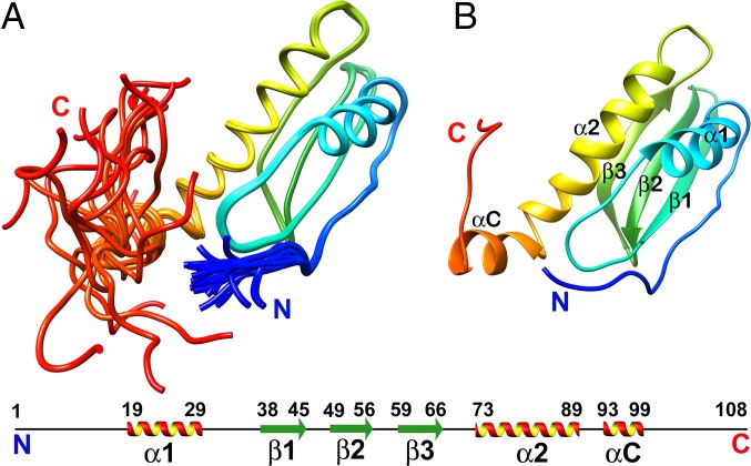
Fig. 3.
Representative structure of W48A/F79A rainbow colored as blue to red from the N to C termini. (A) Ensemble of 20 structures. (B) Secondary structure elements are labeled according to the anticodon-binding domain-like fold, with the additional N and C terminal. The 6xHis-tag at the C terminal of the protein is not shown in the structure.

