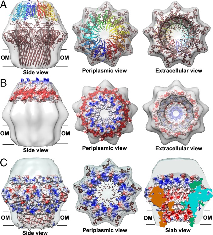
Fig. 5.
Cryo-EM density (EMD ID: 2750) fitting of the CsgG–CsgE complex with the crystal structure of CsgG nonamer (4UV3) and nine copies of the solution NMR structure of double-mutant CsgE W48A/F79A (2NA4). (A) Both CsgE (colored as a blue to red rainbow by models) and CsgG (colored brown) were fitted in the cryo-EM density. Electrostatic potential surfaces of a nonamer of (B) CsgE and (C) CsgG. The surfaces of CsgG in periplasmic view are shown only 20 Å of depth to highlight the interface with CsgE in C. The flexible tail side of CsgE is negatively charged (Fig. 5B and Fig. 4). In contrast, the surfaces of CsgG at the periplasmic vestibule are dominated by positive charges (Fig. 5C and Fig. S8).

