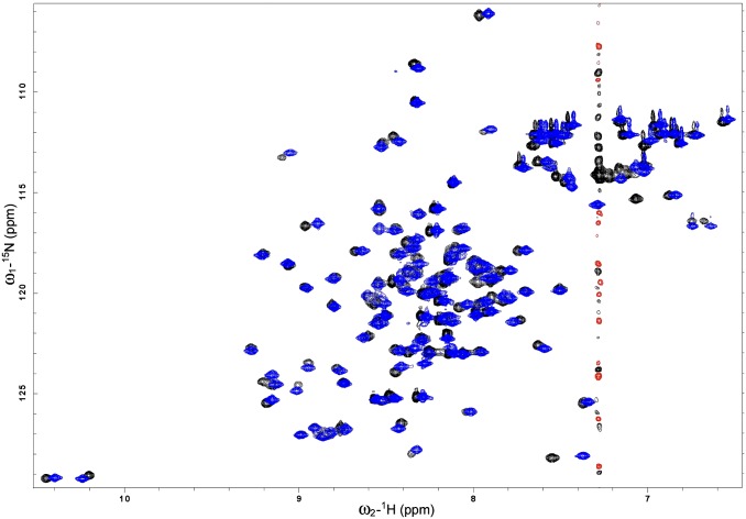
Fig. S5.
Overlay of 2D 1H-15N HSQC spectra of 15N-labeled CsgE W48A/F79A in the presence (black/red) and absence (blue) of 0.5 M Arg. The protein was 100 μM in 10 mM potassium phosphate buffer at pH 5.8, 10% (vol/vol) D2O with or without 0.5 M Arg, 25 °C. The similarity of the spectra suggests that the addition of 0.5 M Arg has no significant effect on the structure of CsgE W48A/F79A.

