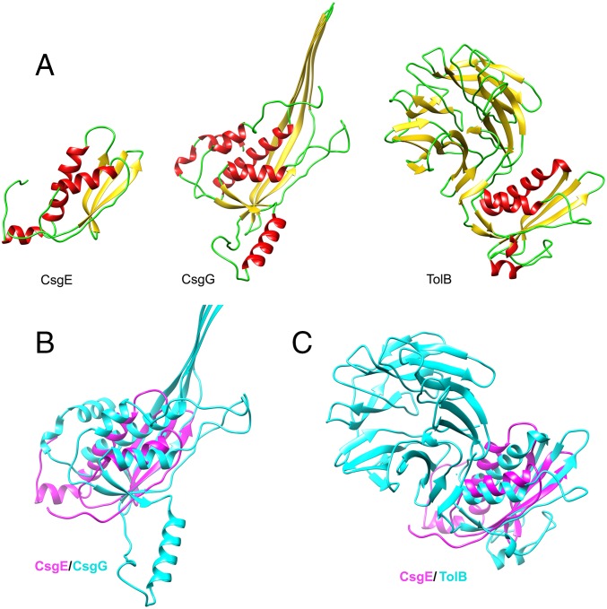
Fig. S7.
Comparison of CsgE with structural homologs with the anticodon-binding domain-like fold. (A) Ribbon diagram for CsgE W48A/F79A, a monomer of CsgG (PDB: 4UV3), and TolB (PDB: 2W8B_A). (B) Overlay of W48A/F79A (magenta) and CsgG (light blue). (C) Overlay of W48A/F79A (magenta) and TolB (light blue).

