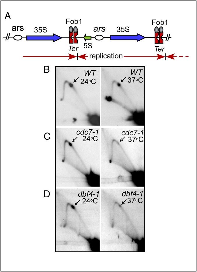
Fig. 1.
Replication termination in the WT and cells with temperature-sensitive mutants of DDK. (A) Schematic diagram of the rDNA repeats showing the locations of the Ter (RFBs) and ars sites. Blue arrows show the direction of rRNA transcripts; red arrows show the direction of replication fork progression. (B–D) Images of Brewer–Fangman 2D gels showing replication fork progression (Y-arc) and the fork arrest at the twin Ter sites (arrows). The cells were grown for 4 h at 24 °C to reach early log phase, transferred to 37 °C for 2–3 h, and then rapidly chilled and treated with Na azide as described in the text and replication intermediates were prepared. Note that whereas fork arrest occurred in the WT cells at both temperatures, it was greatly reduced in the cdc7-1 and dbf4-1 cells at 37 °C.

