
Figure 2. Vole Imaging System.
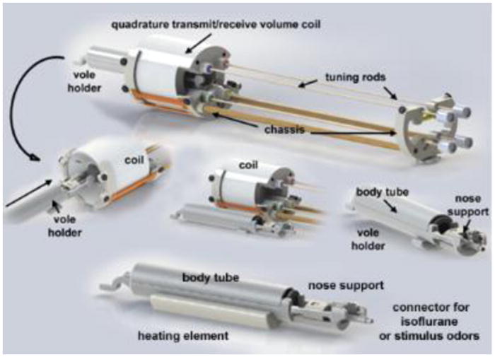
Shown are the different components of the vole imaging system. Note the nose support which includes a hollow tube to secure the front incisors. It is through this tube that odors or volatile anesthetics can be delivered.