
Figure 3. Field of View and Homogeneity.
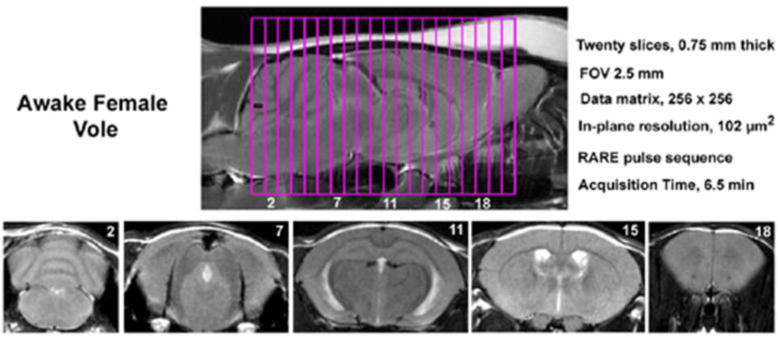
Shown are sagittal and axial views of an awake vole brain. Note the linearity along the Z-axis. The axial images taken from a 20-slice RARE sequence (.75 mm thickness) demonstrate complete brain coverage from the olfactory bulbs to the brainstem. The vole system was provided by Animal Imaging Research, Holden, MA, USA.