
Figure 4. Vole Brain Atlas.
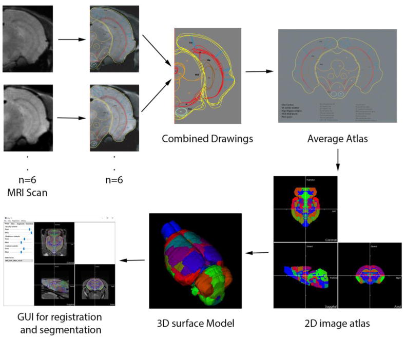
To create an atlas for the vole brain, high resolution MRI scans from 6 voles were collected and registered to standard MRI with affine transform, inspecting and ensuring that boundary surfaces of all the scans matched as perfectly as possible. Separate brain regions were delineated with freehand drawings for each subject. An average scan from all the subjects was created which was used to create a primary template for each slice. The freehand-drawn average atlas was converted to a mathematically robust image format and 3D surface models via an automated 3D segmentation algorithm. This atlas model now can be rotated and deformed to register any MRI image dataset, at which point every voxel in the set of MRI slices is tagged with the fully segmented tissue classification.