Figure 6.


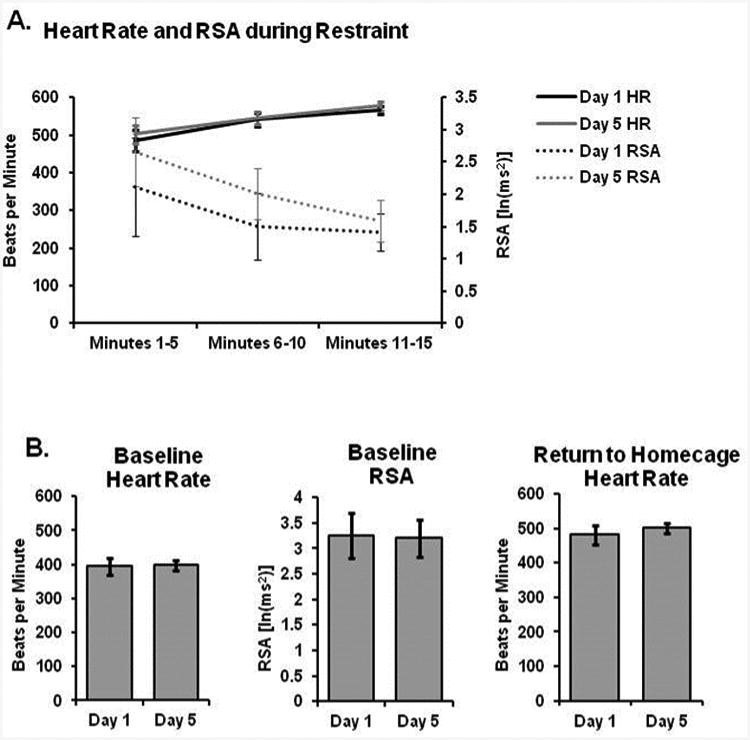
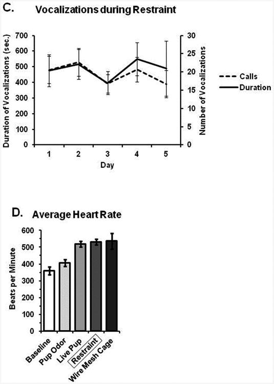
Acclimation to repeated restraint. (A) Heart rate gradually increased while RSA decreased over the course of a 5 minute restraint session; however, there were no differences between days 1 and 5 in either measure. (B) There were no differences in either cardiovascular parameter at baseline, nor following return to the home cage immediately after restraint. (C) Sounds produced during restraint were no different over 5 days of repeated restraint. (D) Average heart rate is shown during restraint in comparison to baseline and various experimental manipulations; heart rate during restraint is roughly similar to that following freely moving exposure to a novel pup or wire mesh floored cage (used for metabolic studies).
