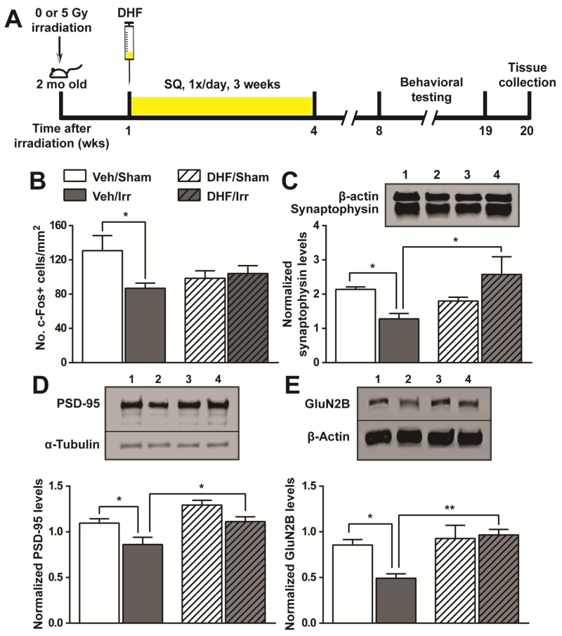
Fig. 4.
Changes in neuronal activities and synaptic proteins following irradiation and DHF treatment. A, experimental timeline highlighting time points of irradiation, DHF treatment, and tissue collection. B, expression of the immediate early gene, c-Fos, upon exposure to a novel environment. The number of c-Fos positive cells in the dentate gyrus was normalized to the area examined. C-E, the levels of synaptophysin (C), PSD-95 (D), and glutamate receptor (GluN2B) (E) in the synaptosomes isolated from the hippocampal formation. Results from two-way ANOVA with Bonferroni’s post-hoc analysis are shown. *, p<0.05; **, p<0.01. N=6 each. Western blot lanes 1-4: Veh/Sham; Veh/Irr; DHF/Sham; DHF/Irr.

