
Figure 2.
Effects of moderate‐intensity continuous training (MICT), and sprint interval training (SIT) on eNOS serine1177 phosphorylation in capillaries and terminal arterioles
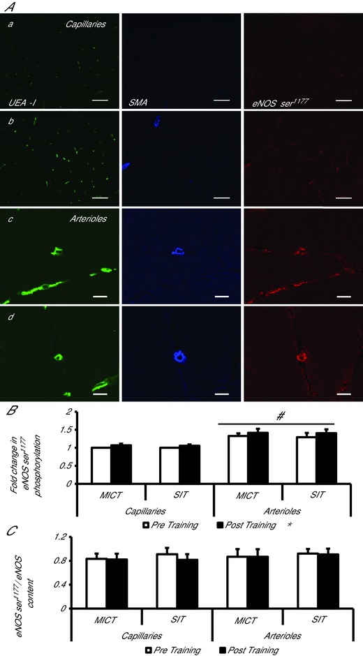
A, representative confocal microscopy images of skeletal muscle from pre‐ (a, c) and post‐training (b, d), in capillaries (a, b) and arterioles (c, d). The skeletal muscle microvascular endothelium was revealed using Ulex europaeus‐FITC conjugated lectin (green). Arterioles and capillaries were differentiated using anti smooth muscle actin in combination with Alexa Fluor 633 conjugated secondary antibody (blue). Skeletal muscle eNOS ser1177 phosphorylation was revealed using Alexa Fluor 546 conjugated secondary antibody (red). Bar represents 50 μm in a and b and 10 μm in c and d. B, mean fluorescence intensity of eNOS ser1177 is summarised (MICT n = 7, HIT n = 8). The mean level of eNOS ser1177 pre‐training was assigned a value of 1, and the relative intensity of eNOS ser1177 post‐training was calculated. C, eNOS ser1177phosphorylation normalised to eNOS content (eNOS content/eNOS ser1177 phosphorylation) (MICT n = 7, HIT n = 8). *P < 0.05, main effect of training. # P < 0.05, main effect of vessel type.